The hip joint is the highest weight-carrying joint in our body. In simple terms, it can be understood as a ball and socket joint which is surrounded by ligaments, muscles, and tendons. The thigh bone or femur joins the pelvis to form the structure of the hip joint.
The joint's ability to carry weight and range of motion can be adversely affected by any severe injury or disease. Mumbai Orthosports is one of the best clinics for Hip Replacement Surgery in Mumbai.
The hip joint consists of:
Given below is a brief explanation of each of these consisting units, based on a discussion with the best Hip Replacement Surgeon in Mumbai.
The hip joint is referred to as the junction where the hip joins the leg to the trunk of the body. It consists of two bones: the thighbone or femur and the pelvis, which is further made up of three bones that are known as ilium, ischium, and pubis.
The femoral head makes the ball of the hip joint while the acetabulum forms the socket. The acetabulum is a deep, round socket that is formed on the outer edge of the pelvis by the fusion of three bones: ilium, ischium, and pubis. The pubis attaches the lower part of the ilium, and the ischium is present at the back of the pubis. The hip becomes stable by the acetabulum, muscles, and ligaments that surround the hip joint.
The Hip Replacement Surgeon in Mumbai further explains that within the acetabulum, the head of the femur rotates and glides. To the acetabulum, a fibrocartilaginous lining called the labrum is attached, which causes the increased depth of the socket.
The femur or the thighbone is the largest bone in the body. Its upper part comprises the femoral head, femoral neck, and the two protrusions called greater and lesser trochanters which serve as sites of muscle attachment. The formation of Hip joint takes place when the head of the femur joins the acetabulum.
The weight-bearing bones of our body are covered by a slippery, white tissue known as Articular cartilage. It is lubricated with a synovial fluid that enables easy and smooth movements of the bones.
Ligaments are fibrous connective tissues that attach one bone to another. Ligaments encircle the hip joint and form a fibrous structure around the joint capsule, thereby making it stable. The ligaments adjoining the hip joint include:
A long tendon called the iliotibial tract runs along the femur from the hip to the knee and serves as a site for attachment of several hip muscles that are described below:
Nerves of the hip carry signals from the brain to the muscles, thereby making the hip movement possible. They also transfer the sensory signals such as pain, temperature, and touch back to the brain. The most important nerves in the hip region are the femoral nerve and sciatic nerve. The femoral nerve is present in the front of the femur, while the sciatic nerve is at the back. There is also an Obturator nerve in the hip, which provides sensory and motor innervation to the thigh.
In addition to these nerves, there are also blood vessels that carry blood to the lower limbs. For example, the femoral artery, one of the longest arteries in the body, arises deep in the pelvis and can be felt in front of the upper thigh.
Hip movements include flexion, extension, adduction, abduction circumduction, and hip rotation. All the anatomical parts of the hip work together to enable these movements. If you have been experiencing problems with hip movements for a long time, you can consult Dr. Abhijeet Savadekar, the best Hip Replacement Surgeon in Mumbai. A Hip Replacement Surgery in Mumbai is a surgical procedure that restores hip mobility by replacing the broken or damaged head of the femur bone with an artificial device. The goal of the surgery is to relieve pain and improve function.

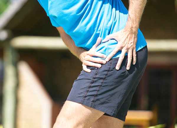
Hip pain, one of the common symptoms that patients complain of, may not always be felt precisely over the hip joint. Pain may be felt in and around the hip joint and the cause for pain is multifactorial.
Know More
Osteoarthritis, also called degenerative joint disease, is the most common form of arthritis. It occurs most often in older people. This disease affects the tissue covering the ends of bones in a joint (cartilage).
Know More
The hip joint is a “ball and socket” joint. The “ball” is the head of the femur, or thighbone, and the “socket” is the cup shaped acetabulum.
Know More
Hip bursitis is a painful condition caused by inflammation of a bursa in the hip. Bursae are fluid- filled sacs present in joints between bone and soft tissue to reduce friction and provide cushioning during movement.
Know More
Femoroacetabular impingement (FAI) is a condition where there is too much friction in the hip joint from bony irregularities causing pain and decreased range of hip motion.
Know More
Hip abductors are a major group of muscles found in the buttocks. It includes the gluteus maximus, gluteus medius, gluteus minimus, and tensor fascia lata muscles.
Know More
Avascular necrosis, also called osteonecrosis, is a condition in which bone death occurs because of inadequate blood supply to it.
Know More
Physical therapy is an exercise program that helps you to improve movement, relieve pain, encourage blood flow for faster healing, and restore your physical function and fitness level.
Know More
The hip joint is one of the body's largest weight-bearing joints and is the point where the thigh bone (femur) and the pelvis (acetabulum) join. It is a ball and socket joint in which the head of the femur is the ball and the pelvic acetabulum forms the socket.
Know More
The hip joint is one of the body's largest weight-bearing joints and is the point where the thigh bone (femur) and the pelvis (acetabulum) join.
Know More
The hip joint is one of the body's largest weight-bearing joints and is the point where the thighbone (femur) and the pelvis (acetabulum) unite.
Know More
The hip joint is a ball and socket joint, where the head of the thighbone (femur) articulates with the cavity (acetabulum) of the pelvic bone.
Know More
 Covid Update